
Application and development of chairside digital systems in implant restorations
2022-06-22
Application and development of chairside digital systems in implant restorations
Since the introduction of computer-aided design and computer-aided manufacture (CAD/CAM) into restorative dentistry and the fabrication of the first CAD/CAM dental restorations, CAD/CAM technology has been developed in dentistry for more than 30 years. The chairside digital system has been upgraded and updated iteratively to enable the design and fabrication of a variety of tooth-supported all-ceramic restorations such as inlays, high inlays, veneers, and full crowns. With the continuous advancement of digital technology and implant technology, the application of chair-side digital technology to achieve implant restorations has become a hot research topic.So far, the more mature chairside CAD/CAM systems include CEREC® (Sirona, Germany) and E4D® (Planmeca, Finland), etc. The CERECAC system was developed by Sirona to support the design and fabrication of implant-supported upper restorations. The chairside CAD software has been upgraded to CEREC version 4.4. The CEREC system is the most commonly used chairside CAD/CAM system in China and the E4D system is also available but does not yet support chairside implant restorations. The CEREC system is the most commonly used clinical study in the literature for chairside digital implant restorations.
Basic process and time efficiency of chair-side digital implant restoration
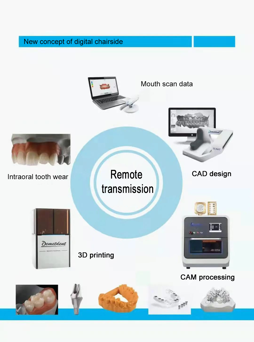
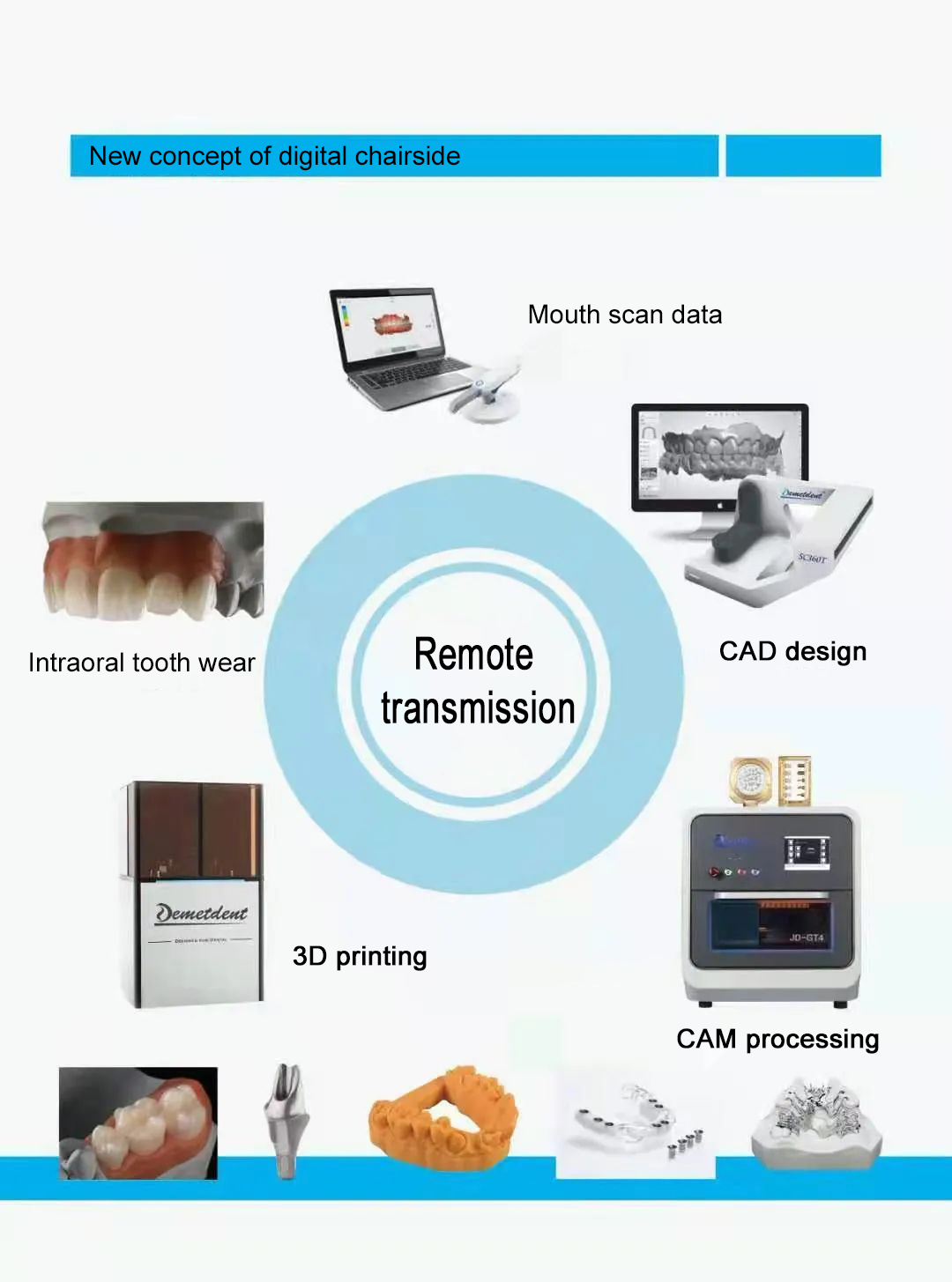
Workflow of chair-side digital implant restoration
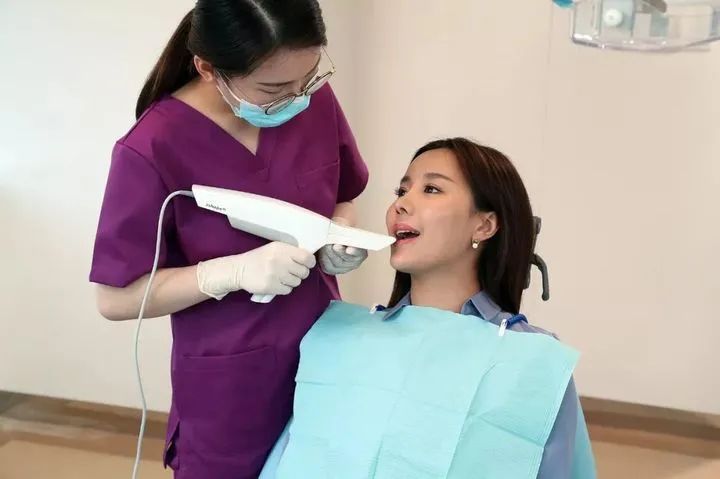
(1) Optical models of the implant position, depth, soft tissue, adjacent teeth, opposing teeth and occlusal records were obtained by intraoral scanning.(2) The acquired optical information is transmitted to the computer and the CAD software is used to design the upper restoration.
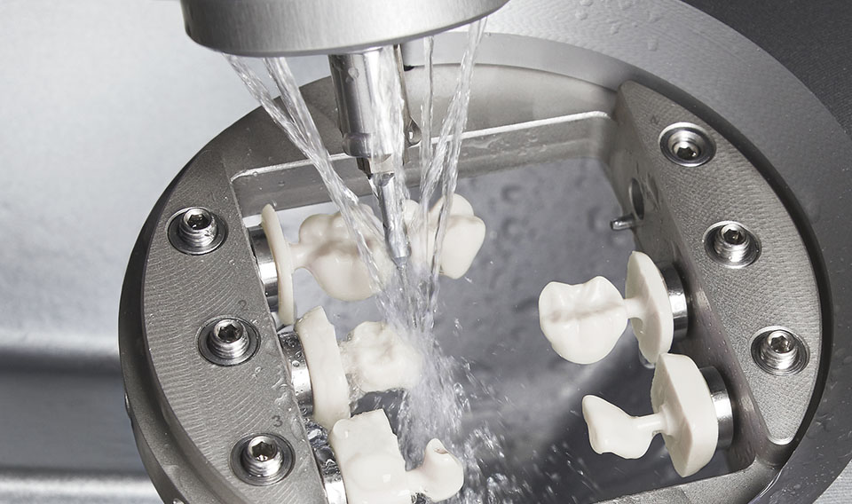
(3) The designed restoration solution is transmitted to the grinding and cutting equipment for grinding and cutting, and after completion of grinding and cutting, it is burnished and polished, and finally, the clinical intraoral trial is performed. Joda et al. studied and analyzed the efficiency of the traditional method and digital method techniques applied to the single posterior dental implant restoration process. It was shown that the digital process took less time and was more efficient than the traditional method for fabricating implant-supported fixed crown restorations. Another study by Joda et al. compared the entire process of individualized titanium abutment + CAD/CAM fabricated zirconia superstructure with standard titanium abutment + porcelain crown restoration after a single missing posterior tooth. The study showed that the digital process resulted in a good intraoral fit with little or no adjustment. The digital process produces implant-supported restorations with chairside adjustments in 1/3 the time of conventional methods.
Materials that can be chair-worked
All-ceramic restorative materials: Feldspar-based mainly include Vitablocs series (Vita, Germany) and CERECBlocs series (Sirona, Germany). Glass ceramics mainly include mica-based, leucogranite-based and lithium silicate-based glass-ceramics. The white garnet-based glass-ceramics products are IPSEmpressCAD and IPSProCAD (Ivoclar Vivadent, Liechtenstein). Lithium silicate-based glass-ceramics are available as IPSe.maxCAD (Ivoclar Vivadent, Liechtenstein). Alumina ceramics are Procera All Ceram (Nobel Biocare, Sweden), and similar products are available from Vita and Sirona. The main glass-permeable ceramics are In-Ceram spinel (Vita, Germany); in addition, there are zirconia all-ceramic materials. informs (Sirona, Germany), a translucent zirconia porcelain block available for the CEREC system, was launched in 2011 and can be used for zirconia all-ceramic crowns, bridges and individualized implant abutments. For several years, Lava Ultimate Restorative (3MESPE, Germany) CAD/CAM material has also emerged as an ideal material for implant-supported crown restorations.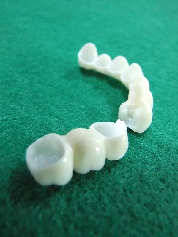
Resin materials: PMMA (polymethylmethacrylate) resins and composite resins are the most commonly used materials for temporary restorations. CAD/CAM can increase the strength of temporary crowns. One of the representatives of CAD / CAM temporary restorative materials is TelioCAD (Ivoclar Vivadent, Liechtenstein), which is made of glued PMMA blocks prefabricated with a high degree of fracture resistance. PMMA blocks are pre-polymerized to provide greater strength and homogeneity, unlike conventional PMMA materials, without polymerization shrinkage. Can be used to create temporary crowns, bridges and temporary abutments.
Performance of Chairside CAD/CAM Machinable Materials: An in vitro study comparing the fracture strength of temporarily fixed bridge restorations made by conventional methods with those made by CAD/CAM found that CAD/CAM fixed bridges were stronger. However, the authors note that this finding is controversial when applied to interpret the material when subjected to occlusal forces intraorally. Stawarczyk et al. studied the rate of autogenous wear of several CAD/CAM resin blocks (ZENOPMMA, artBlocTemp, TelioCAD, CAD-Temp) in relation to the rate of wear on maxillary enamel and compared it with VITAMARKII (Vita ) and artificial polymeric resins, and found that both artificial polymeric resins and CAD/CAM resins had lower wear rates on the maxillary enamel than glass ceramics and that the CAD/CAM resin block had a lower autogenous wear rate than the artificial polymeric resins.
Accuracy
Ganz et al. considered an acceptable margin gap of 20-70 μm for implant-supported restorations before bonding. With the development of digital technology, the precision of CAD/CAM restorations has improved. The restorations produced by this process were found to meet clinical requirements and were more accurate than those produced by conventional methods. The accuracy of the structure of the resin material as a tooth-supported upper restoration has been well studied. Cases have been reported using CEREC Paradigm MZ100 crowns for implant-supported provisional restorations, but the accuracy and marginal clearance of implant-supported provisional restorations made with CAD/CAM resin materials have not been reported in the literature. A 3D digital model is obtained from a chair-side intraoral scan by combining numerous 3D images obtained by scanning the same part from different angles.The combination of these 3D images inevitably introduces an alignment error. The magnitude of this error is determined by the scanning technique and the alignment algorithm. The source of scanning error is different for digital implant restorations compared to the fabrication of digital conventional restorations. The irregular and variable nature of the natural tooth preparation surface increases the difficulty of intraoral scanning, and the subsequent processing of the image and noise cancellation can cause loss of marginal detail. Unlike implant-supported restorations, which have a smooth implant surface with specific and regular geometry and 3D dimensions, the scan is aligned with the software's built-in scan body database morphology to determine the 3D position of the implant.
The accuracy of the intraoral scan consists of two parts: firstly, the accuracy of the scan of the micro-geometry, which is mainly reflected in the scan of the individual implants. The second is the accuracy of the transverse arc of the multi-unit system, which is mainly reflected in the scanning of the multi-unit system. It has been shown that there is no significant difference in accuracy between intraoral scanning and conventional impression methods in laboratory studies of the transfer of single implants to superior three-dimensional positions, both within clinically acceptable ranges. Vander Meer et al. investigated the accuracy of three intraoral scanners, CEREC, iTero and LavaCOSTM, for post-implant transfer of implant 3D positions. The results of the study suggested that all three intraoral scanners had small angular errors. Compared to the three, LavaCOSTM had the smallest distal error and the best stability, while CEREC had the largest distal error and the least stability. Andriessen et al. investigated the accuracy of intraoral scanning applied to multiple implant scanning in the mandibular edentulous jaw. Both the distance error and the angular error were too large resulting in the inability to create superstructures with a good fit.
The current technique for scanning multi-implanted and edentulous jaws mainly relies on scanning the model after conventional impression taking to obtain digital data. The current CEREC chairside system uses the Biogeneric bio-reconstruction design software to reconstruct the occlusal surface based on the patient's dental morphology with reference to the contralateral jaws. This technique also has an expected error of 0.5 mm in the occlusal surface, which requires manual adjustment. The CAD software design for implant restorations includes the entire crown morphology and gingival penetration profile, which is more computationally intensive and difficult than for full-crown restorations. The fineness of computer-aided manufacturing (CAM) restorations depends on the smallest turning pin diameter (approximately 1 mm), and smaller restorations cannot be represented in detail. Since the tissue surfaces of implant restorations are smooth and continuous, no sharp edges are encountered during cutting. The diameter of the restoration surface detail is larger than the minimum diameter of the tuning pin, thus ensuring accurate and reliable precision of the tissue surface.
Application and development of chair-side digital systems in implant restoration
Implant support applied to single tooth loss
Superstructure restoration In 2004, Fritzsche et al. used CEREC to fabricate implant-supported super crown structures for single missing teeth. The two main retention methods for implant-supported single crowns are cemented and screw-retained. The fabrication of cement-retained full-crown restorations on implant abutments by digital methods has been reported several times. Joda reported a case of a fully digital process for fabricating an implant-supported posterior crown on an abutment using iTero intraoral scanning and digital software to design a fully anatomical screw-retained RNC crown, which was obtained directly with CAD/CAM cutting.Joda concluded that a fully digital process for fabricating RNC fully anatomic implant-supported crowns is fully feasible. The authors also compared the intraoral aesthetics of the one-piece abutment crown with the bonding of preformed and individualized titanium abutments. The use of prefabricated titanium abutments was found to be superior to personalized abutments in terms of aesthetic results and overall treatment time (clinical time + technician's office time). I resulted from the studio of Jiehua Tian et al., who were the first in China to report the use of the CEREC chairside scanning digital processing technology for immediate restorations in the aesthetic area, suggest that using the digital chairside system for immediate single-visit implant restorations is a feasible clinical procedure that can significantly reduce clinical restoration time, especially the restoration set-up time, with high patient satisfaction and good results during the observation period of 3-6 months. The clinical results were achieved within 3-6 months of observation. The abutment is a unique structure in implant restoration, and its shape, strength and colour are related to the clinical results of the implant restoration. The design of individualized abutments to meet the needs of different patients has always been a concern of the implant restoration profession.
Currently, chair-side digital prosthetic devices allow for the fabrication of individualized abutments. Bernardo et al. reported a case of a single maxillary premolar tooth loss in a single visit with immediate restoration of a CAD/CAM zirconia abutment + all-ceramic crown after implant placement. Subsequently, Bonaudo et al. reported an immediate chairside restoration of a single mandibular premolar with a single visit, and Gougaloff et al. performed an immediate chairside implant restoration using CEREC, making a conventional impression followed by scanning of the model and fabrication of a provisional crown on CEREC software. with temporary crowns as implant immediate restorations to guide soft tissue healing.
In 2016, Proussaefs used the same method to fabricate screw-retained temporary PMMA crowns for immediate restorations. All of the above reports were model scans followed by fabrication of temporary restorations for immediate restorations in the mechanic's office. The use of chair-side intraoral scanning to design and fabricate provisional restorations will also be a trend. Soft tissue management in aesthetic area implant restorations is related to the final outcome of the restoration. The use of digital technology in implant restorations also offers new methods of soft tissue processing and shaping in the aesthetic area. When the temporary restoration is removed, the soft tissue collapse makes it difficult to accurately replicate the soft tissue around the implant with conventional impressions. To address the problem of soft tissue collapse during intraoral scanning after removal of the provisional crown, Joda utilizes the "Personalized Scanning Body Technique" to prevent soft tissue collapse during intraoral scanning in the upper part of the implant, which is prone to change. The predictability of the soft tissue morphology was increased and good aesthetic results were obtained. However, the entire submucosal gingival cuff shape is not yet accessible, so control of the penetrating gingival contour is still lacking.
A similar technique was reported by Proussaefs et al. Joda reported a new digital method for soft tissue shaping of the upper part of the implant: by combining digital thin-layer CBCT with the STL file obtained after intraoral scanning, a personalized healing abutment was visualized on the software and Cutting PMMA was obtained and the soft tissue morphology was plasticized during the same period of osseointegration.
Applications for implant-supported superstructure restoration in multiple missing teeth and superstructure restoration in edentulous jaws:
Because the accuracy of chair-side intraoral scanning decreases as the scanning area expands, it is currently not sufficient to meet the clinical requirements for fabricating superstructures for multi-tooth contiguous loss and edentulous implant restorations.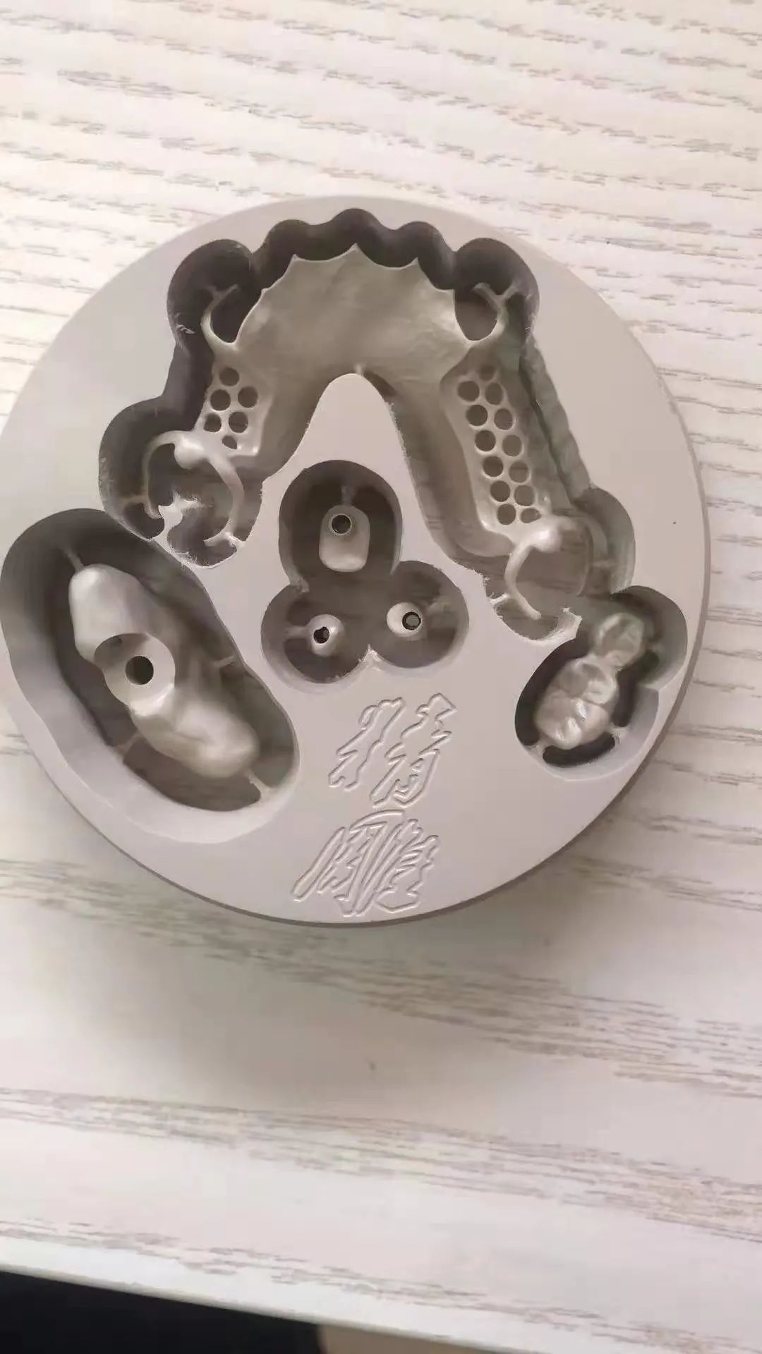
The Future of Chairside Digital Implant Restoration
With the increasing ease of intraoral scanning, computer-aided manufacturing and processing technology, the increased compatibility and openness of the data and equipment of the various digital systems, and the emergence of new cuttable materials, it helps to contribute to the trend of fully digital realization of implant restorations. The chair-side digital technology has been successful in single-tooth implant restorations, and more chair-side digital technologies will be used for clinical research and exploration of implant restorations in the future: chair-side digital restoration of single-tooth implants in the posterior region, chair-side digital restoration of immediate implants in the aesthetic region, chair-side digital technology for personalized abutment fabrication, etc. have been reported in the literature and require long-term clinical observation with large sample sizes; the use of chair-side digital technology The use of chair-side digital technology for diverse peri-implant soft tissue shaping is a predictable trend in the clinical application of chair-side digital technology in the field of implant restoration at this stage. For the chair-side digitalization of edentulous implant restorations, a longer period of multidisciplinary and multidisciplinary exploration is needed before it can be realized. CNC milling machine operation need to pay attention to what?
 - What is orography?
- What is a denture?
- What is a CNC milling machine?
 Oral chairside device prosthetic system industry development process3
 - Development process of the prosthetic systems pipeline for chairside oral devices-two
 Oral chairside device restorative system industry development process one
  CNC milling machine on the teeth: grinding machine
  ## what's a dental milling machine?
  Your satisfaction is our ultimate goal